The cord below the break
Epidural stimulation of the spinal cord does not drive paralysed muscles. It stimulates sensory fibres, biases motoneurons toward threshold, and lets a surviving descending command become effective again. Why that distinction decides everything, and why the recovered circuit is not the original one.
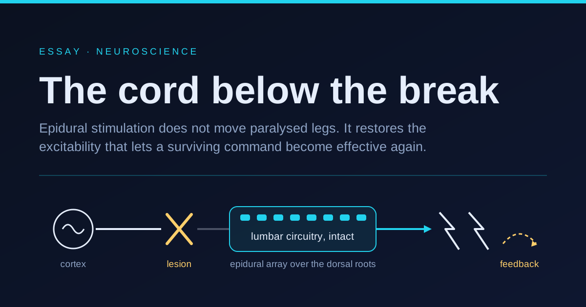
A paddle electrode for the lumbar spinal cord carries sixteen contacts on a strip of silicone roughly the size of a stick of chewing gum. It sits in the epidural space, on the dorsal side, inside the bony canal but outside the dura. It touches no neuron. Switch it on in someone whose legs have not moved voluntarily for years and, at the settings that matter clinically, nothing that looks like walking happens.
What happens is stranger and much more useful. The legs become recruitable. A command that had been arriving at the lumbar cord for years without visible effect starts producing movement. The device is not driving the muscles. It is changing what the cord does with instructions it was already receiving.
That distinction is the entire subject, and almost every popular account of these results gets it backwards.
A controller, not a cable
The everyday mental model of the spinal cord is a wiring loom: the brain decides, the cord conducts, the muscles obey. Cut the loom and the signal stops. Under that model, restoring movement means restoring conduction, which means regenerating axons across the lesion, which is a problem nobody has solved in humans.
The cord does not work that way. It holds on the order of ten million neurons, the overwhelming majority of which are interneurons that never leave it. Each corticospinal tract contributes roughly a million axons descending into that population, and most of them do not synapse onto motoneurons at all. They terminate on interneuronal networks that already know how to do things. Load compensation, reciprocal inhibition between flexors and extensors, the reflexive extension that stops a leg from folding when weight lands on it: all of that is computed locally, on a millisecond timescale that no descending command could meet.
In cats, a cord surgically isolated from the brain will still generate alternating stepping on a treadmill, the classic evidence for a central pattern generator. In humans the case for an autonomous rhythm generator is thinner, and honest summaries stop at a weaker claim: the human lumbar cord contains powerful circuitry organised around proprioceptive feedback, whether or not it can produce a rhythm with no input at all.
So a lesion at, say, the eighth thoracic segment does not destroy the machinery for walking. It orphans it. Everything below the break is intact, wired, and starved of the descending drive that normally holds it near its operating point. Motoneurons sit chronically hyperpolarised, several millivolts too far from firing threshold, and any residual command that trickles across the lesion arrives too weak to close the gap.
What the current actually touches
Put an electrode in the epidural space and the nearest excitable structures are not motoneurons, which sit deep in the ventral horn behind the whole cross section of the cord. They are the dorsal roots, the sensory bundles entering the cord from the periphery, curving right past the electrode.
Two facts then decide everything. Activation threshold falls steeply with axon diameter, roughly as the inverse square, so the fattest fibres go first. And the fattest fibres in the dorsal root are the group Ia and Ib proprioceptive afferents, twelve to twenty micrometres across, carrying the muscle spindle and tendon organ signals that report length and force.
Epidural electrical stimulation is therefore a sensory technique wearing a motor costume. Each pulse fires a synchronous volley of proprioceptive afferents, which excite motoneurons through the same monosynaptic and polysynaptic paths a tendon tap uses. You are not pressing the accelerator. You are pressing the muscle’s own report of its own stretch back into the cord, forty times a second, and letting the existing reflex architecture do what it would have done with that information anyway.
The consequence is a bias, not a command. Write the condition for movement crudely: a motoneuron fires when the residual descending drive plus the stimulation-induced depolarisation exceeds threshold. Stimulation supplies the second term. If it is too small, nothing changes. If it is too large, the muscles contract on their own and the person is a passenger. The therapeutic window is the band where the sum crosses threshold only when the person intends it to, and finding that band for each muscle group in each individual is most of what the clinical work consists of.
This also explains why so many injuries classified as motor complete respond at all. Complete is a bedside classification, not an anatomical one. Post mortem and electrophysiological studies keep finding surviving axons crossing lesions in people who had no detectable voluntary movement, a condition sometimes called discomplete. Those spared fibres were never useless. They were subthreshold.
The reason timing beats amplitude
If stimulation simply added excitability, more of it would be better. It is not, and the reason is a small piece of axonal physics with large clinical consequences.
An action potential fired in the middle of an axon propagates in both directions. The orthodromic half travels into the cord and does the useful work. The antidromic half travels back out toward the muscle, and when it meets a genuine spike coming the other way from a spindle, the two collide and both are extinguished, because the membrane behind each is refractory. Continuous stimulation at forty pulses per second therefore does not merely add a signal. It systematically deletes the natural proprioceptive traffic on the very fibres it is exciting.
That is a self defeating design. The cord’s whole competence rests on knowing limb position and load, and the stimulator has been erasing that knowledge in exchange for a crude tonic boost.
The fix is spatiotemporal: map each electrode subset to the muscle groups it recruits most selectively, then fire it only in short bursts, only during the phase of the step cycle in which that group is supposed to act, and leave the fibres quiet the rest of the time. This is the change that separates the modern results from two decades of ambiguous tonic stimulation studies. It is also the reason the implant needs to know where in the gait cycle the person currently is, which is what drags the rest of the system into existence.
Closing the loop from the cortex
Once the stimulator has to be phase locked to intention, the obvious question is where the phase information comes from. Motion sensors work for locomotion. Reading the cortex directly works for everything else.
The brain to spine bridge built at Lausanne does that. Cortical activity is recorded epidurally over the sensorimotor cortex, decoded into an intended movement, and the prediction is used to select and modulate the spinal stimulation pattern. The result, published in Nature in 2023, was a participant with chronic tetraplegia walking with a natural gait under volitional control, on terrain including stairs.
The system architecture is the interesting part. A decoder that had to specify muscle activations directly would need to output tens of continuously varying channels, and no practical cortical recording gives you that reliably for years. This decoder does not have to. It only needs to identify which of a small library of elementary movements the person is trying to make. The cord expands that into the actual pattern of activation, with all the load compensation and interlimb coordination it already knows. Low bandwidth intention in, high bandwidth coordination out.
The neurons that were not needed before
The most disorienting finding in this literature is not about hardware.
After stimulation combined with intensive rehabilitation restored walking in nine people with chronic injury, the Lausanne group went looking for the cellular substrate in mice, using single nucleus RNA sequencing and spatial transcriptomics to build a molecular map of recovery. Reported in Nature in 2022, one population stood out: excitatory lumbar interneurons expressing the transcription factor Vsx2. Silencing them abolished the recovery. Activating them reproduced it. And in uninjured animals, silencing them did essentially nothing, because walking never depended on them in the first place.
Read that again slowly. The circuit that recovers is not the circuit that was lost. Rehabilitation with stimulation does not repair the original pathway; it recruits a population that was previously along for the ride and reorganises the cord around it. Whatever this therapy is doing, restoration is the wrong word for it.
Where this stands in 2026
The bridge has moved up the cord. A digital bridge between the brain and the cervical spinal cord has been implanted in three people with chronic incomplete tetraplegia, using a library of stimulation patterns to build up control of elementary arm and hand movements, with the system reported stable beyond a year. Arm and hand are much harder than legs, for reasons discussed below, and this is the first credible demonstration that the same architecture transfers.
A parallel line combines an intracortical brain computer interface with patterned stimulation of both the spinal cord and the cortex. Reported in Nature Medicine in 2026 in a person with complete injury, it produced real time control of the person’s own hand and, more importantly, gains in movement and sensation that persisted with the system switched off. That carryover is the signature of reorganisation rather than substitution.
The non invasive route has reached the market. Transcutaneous stimulation over the cervical spine, which recruits the same afferents at much higher current through skin and bone, was tested for hand strength and function in chronic tetraplegia and reported in Nature Medicine in 2024. The device received de novo authorisation in the United States in December 2024, and clearance for home use in November 2025. No surgery, smaller effects, a far larger eligible population.
Replication outside the two founding laboratories is beginning, which matters more than any single headline. A 2026 pilot cohort reported long term functional recovery and immediate neuromuscular facilitation after epidural stimulation in chronic injury, and a 2025 case study extended the approach to a lesion of the conus medullaris, where the target circuitry is itself partly damaged.
What breaks
Everything above rests on small, unblinded, single arm studies. Nine participants is a landmark in this field and a rounding error anywhere else. There is no sham arm, and there cannot easily be one, because participants know when a stimulator is on. The intervention is inseparable from months of intensive supervised rehabilitation, and untangling the contribution of each is not something any published trial has done.
Responder rates need reading with care. Headline figures like nine in ten participants improving come from single arm trials against a within subject baseline, with the improvement threshold defined in advance by the people running the study. That is a legitimate design for a first trial and a weak basis for comparing therapies.
The mechanism sets a hard eligibility limit. If stimulation works by biasing residual descending drive toward threshold, then an injury with no surviving axons has nothing to bias. Some people will get standing and stepping in a harness that never becomes household mobility.
Hands are the real wall. Locomotion is rhythmic, bilateral, low dimensional, and supported by circuitry evolved to run semi autonomously. Grasping is aperiodic, high dimensional, and has no pattern generator to lean on. The cervical cord will not expand a coarse intention into a functional grasp the way the lumbar cord expands one into a step, which is why the arm and hand work reports elementary movements rather than restored dexterity.
And the Vsx2 result, the most conceptually important piece of the whole story, is mouse work. No one has shown that the human cord reorganises around the homologous population, and there is currently no way to look.
Further reading
- Walking naturally after spinal cord injury using a brain to spine interface, Nature, 2023
- The neurons that restore walking after paralysis, Nature, 2022
- Non invasive spinal cord electrical stimulation for arm and hand function in chronic tetraplegia, Nature Medicine, 2024
- A neuroprosthesis for restoring hand movement and sensation in a person with complete tetraplegia, Nature Medicine, 2026