Speaking to the retina in the wrong language
Restoring sight is not a pixel count problem. The optic nerve carries around forty parallel feature streams rather than an image, and every visual prosthesis is a decision about how much of that native encoding to keep. What the 2026 clinical results actually establish, and what they leave untouched.
A person in their eighties, blind in the centre of the visual field for years after geographic atrophy destroyed the photoreceptors of the macula, sits in a clinic and reads a word off a chart. The reading is slow. It is monochrome. It is done with a two millimetre chip carrying 378 pixels, sitting under what remains of the retina, powered by infrared light thrown at it by a pair of glasses.
The obvious way to read that scene is as a resolution story: 378 pixels today, more tomorrow, and eventually enough for something like sight. That reading is wrong, and the reason it is wrong says more about the eye than about the device.
The retina does not send pictures anywhere. By the time a signal leaves the eye it has already been taken apart, and any implant that intervenes is not filling in missing pixels. It is interrupting a translation midway and hoping the rest of the sentence still parses.
What the optic nerve actually carries
Start with the compression. A human retina holds roughly 120 million rods and 6 million cones. The optic nerve that leaves it carries roughly one million axons. Two orders of magnitude go missing between the surface that catches photons and the cable that reports on them.
That reduction is not a quality setting. Between the photoreceptors and the exit lie bipolar, horizontal and amacrine cells, arranged into circuits that decompose the image into parallel streams. Each stream is carried by its own class of retinal ganglion cell, and each class tiles the whole visual field independently, so the same patch of world is reported several dozen times over, each time filtered for a different property.
The catalogue is best characterised in mouse, where anatomical, functional and transcriptomic evidence converges on something in the region of forty ganglion cell types. Primate retina is less exhaustively mapped but shows the same architecture: midget cells carrying fine spatial detail and colour opponency, parasol cells carrying motion and flicker, and a long tail of smaller populations. The organising principle, described decades ago and still the right frame, is that parallel information processing channels are created in the retina itself rather than downstream.
So a ganglion cell is not a pixel. It is a feature detector with a specific polarity, a specific latency, a specific receptive field size and a specific downstream target. Two adjacent cells in the same square of retina may be reporting on entirely different things. The optic nerve is closer to forty superimposed edited video feeds than to one raw one.
This is the fact that sets the terms for every prosthesis. Electrical stimulation is indiscriminate. A current pulse recruits whatever excitable membrane sits nearest the electrode, with no regard for which of the forty dialects that membrane speaks.
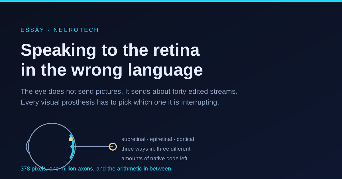
Three places to cut in, and what each one costs
The visual pathway offers a choice of insertion points, and the choice is really a choice about how much of the native encoding you are willing to throw away.
Cut in under the retina, where the photoreceptors used to be, and you keep almost everything. Your electrodes drive the bipolar cells, and the remaining inner retinal circuitry performs its own decomposition on whatever you hand it. You are not obliged to know the code, because the code is still running. This is the subretinal approach, and the photovoltaic version of it is elegant: each pixel is a tiny solar cell, so no wires cross the eye wall and no external electronics need to be implanted. Glasses capture the scene, process it, and project it back onto the chip in near infrared, invisible to any surviving photoreceptors and bright enough to drive current without heating tissue.
Cut in on top of the retina and you address ganglion cells directly. In principle this is the highest fidelity option, because you are talking to the output layer itself. In practice the ganglion cell axons run across the retinal surface in bundles on their way to the optic disc, so an electrode sitting above them stimulates axons of passage belonging to cells located far away. Patients report elongated streaks rather than dots, oriented along the nerve fibre paths. The first commercial epiretinal device is no longer manufactured.
Cut in at the visual cortex and you skip the eye entirely, which is the only option when the optic nerve is gone. You also throw away the entire retinal encoding and must synthesise something the cortex will accept. What stimulation produces there is a phosphene, a spot of apparent light, and the open question is whether a field of phosphenes can be assembled into form.
The arithmetic in the right panel is worth doing slowly, because it is the thing that bounds the whole enterprise. One degree of visual angle projects to about 288 micrometres on the human retina. A chip with a pitch of 100 micrometres therefore gives each pixel an angular footprint of about 21 arcminutes. Normal acuity resolves one arcminute, which is what 20/20 means. Twenty one arcminutes puts the ceiling somewhere near 20/420. Put less formally: each pixel spans about the width of a pencil held at arm’s length, and nothing downstream can invent detail finer than the grid that produced it.
The obvious response is to shrink the pitch, and the obvious response fails for reasons of electrochemistry. Safe charge injection scales with electrode surface area, so a smaller pixel can deliver less charge before the electrode begins to corrode or the tissue begins to suffer. Meanwhile the current still spreads through conductive tissue at roughly the same radius. Past a certain point, tightening the grid simply makes adjacent pixels stimulate each other’s targets, and effective resolution stops improving while nominal resolution keeps rising.
Where this stands in 2026
The subretinal photovoltaic approach produced the year’s substantive clinical result. A trial of 38 participants with geographic atrophy secondary to age related macular degeneration reported twelve month outcomes in the New England Journal of Medicine: a mean improvement of 25.5 letters on the standard chart, with a large majority of participants able to read letters, numbers and words that they could not read before. In July 2026 the manufacturer announced European commercial launch after CE marking, which as far as form vision is concerned makes this the first such device to reach a market rather than a trial.
On the cortical side, the notable 2025 result is quieter and more foundational. A team implanted 100 electrode arrays in two totally blind participants and recorded cortical activity while stimulating, over six months. The finding is that neural activity in visual cortex predicts phosphene threshold and brightness, which matters because it is the precondition for a closed loop device: if you can read out what a stimulus actually produced, you can correct the next one. Cortical prostheses otherwise remain investigational, and the state of that field is still one of small cohorts and single digit numbers of implanted participants.
A third line avoids electrodes altogether. Optogenetic therapy delivers a light sensitive channel protein, typically a red shifted channelrhodopsin, into surviving ganglion cells by viral vector, then drives them with goggles that convert the scene into light at the right wavelength and intensity. The single patient result published in 2021 remains the landmark, and the current state of the approach is cautious optimism with thin cohort level data. Its structural advantage is that resolution is set by which cells express the protein rather than by electrode geometry. Its structural problem is the same one as everywhere else: expression is not type specific, so every ganglion cell that takes up the vector is driven with the same signal regardless of what it normally reports.
What this does not prove
Twenty five letters of improvement is a real clinical effect and should not be deflated. It should also not be over read.
The reading is assisted. The glasses in these systems apply digital zoom, and some of the letter recognition is magnification rather than implant acuity. That is a legitimate engineering solution to a legitimate problem, but it means the reported acuity and the chip’s optical resolution are not the same number.
The vision is monochrome and effortful, and participants in these trials keep their peripheral vision, since geographic atrophy spares it. So the device restores a small central island of form vision that has to be integrated with intact surrounding vision, a rather different perceptual task from restoring sight to someone with none. Whether the same approach transfers to complete blindness is not established by this trial.
Twelve months is short for an implant that has to survive under a retina for decades, and the procedure is retinal surgery with the complication profile of retinal surgery.
Underneath all of it sits the encoding problem, which none of these approaches solves. Every one of them drives a population of cells with a signal that ignores the population’s internal structure. A patch of retina asked to report an edge, a direction of motion, a colour opponency and an increment simultaneously receives, instead, one undifferentiated pulse. The most demanding research programme in the field takes this seriously and tries to identify individual ganglion cells by their electrical signatures on a dense array, classify them by type, and then stimulate each one with the pattern appropriate to its own dialect. That would be speaking the language rather than shouting at it. It is also, at present, a laboratory technique operating on isolated retina, not a clinical device.
Which leaves an uncomfortable observation about why any of this works at all. It works partly because the brain is doing most of the interpretive labour. Patients improve over months of use with the hardware unchanged. They are learning to read a signal that was never designed to be readable, in much the way that a listener adapts to a heavy accent. That plasticity is the field’s quiet dependency, and it is also the reason to be careful about attributing outcomes to devices.
Restoring vision, in the sense the phrase suggests, is not what is happening. What is happening is that a channel is being opened into a system that still knows how to see, and the system is meeting it more than halfway.
Further reading
- Subretinal photovoltaic implant to restore vision in geographic atrophy due to AMD, the twelve month trial report.
- Neural correlates of phosphene perception in blind individuals, on reading out what cortical stimulation actually produced.
- The types of retinal ganglion cells: current status and implications for neuronal classification, for the parallel channel architecture.
- Brain machine interfaces for vision restoration: the current state of cortical visual prosthetics, a survey of the cortical route.