Sound into the skull
Low intensity focused ultrasound can put mechanical energy into a target sixty millimetres deep without an incision, and implanted electrodes have now recorded the effect from inside that target. The physics of why sound gets there when fields cannot, why the bone is the hard part, and why nobody can yet say what the sound does to a neuron.
A deep brain stimulation electrode aimed at the subthalamic nucleus has to cross roughly 80 millimetres of living tissue to reach a structure about 8 millimetres long. Getting there costs a burr hole, a planned trajectory that threads between vessels and ventricles, an implanted pulse generator, and months of programming afterwards. It works, well enough that it has been standard care for Parkinson’s disease for a quarter of a century. It also carries the risks that any permanent hardware in the head carries, which is why the eligible population is a small fraction of the population that might benefit.
So the obvious question is why anyone has to drill. The scalp is right there. Put electrodes or a coil on the outside and aim inward.
The answer is that you cannot, and the reason is not engineering timidity.
Why sound goes where fields cannot
Transcranial magnetic stimulation drives a coil at kilohertz frequencies. At those frequencies the head sits deep inside the coil’s near field, where the induced electric field is not a beam that can be pointed but a diffuse function of geometry that decays monotonically away from the winding. Every design that pushes a stronger field into deep structures pushes a stronger field through everything above them first. Quasi-static fields sourced from outside a volume do not have interior maxima. Clever schemes exist, temporal interference between two high frequency carriers being the most discussed, and they buy real depth, but they are working against the same monotonic decay rather than around it.
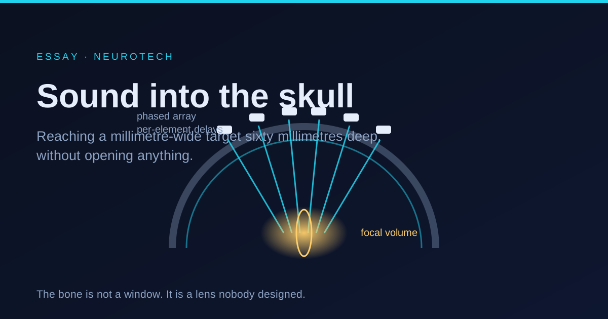
Sound obeys a different rule, because at the relevant frequencies it is a wave in the geometric sense. At 500 kHz the wavelength in soft tissue is about 3.1 millimetres, since the speed of sound there is close to 1540 metres per second. Three millimetres is small compared to a head, and that ratio is what makes focusing possible: a curved aperture, or an array whose elements are fired with individually chosen delays, can arrange for every path to arrive in phase at one chosen point and out of phase everywhere else. The energy is not strongest at the surface. It is strongest where the arithmetic says it should be.
The focus is not a point. Diffraction sets its size, and the useful approximation is that the lateral width scales with the wavelength multiplied by the f-number of the aperture, while the axial length scales with the wavelength multiplied by roughly the square of the f-number. For a 500 kHz array with an f-number near 1, that means something like 3 millimetres across and closer to 20 millimetres along the beam axis. The focal volume is a cigar, not a marble, and its long axis is the most under-discussed limitation in the entire field. A target 8 millimetres long sits inside a spot two and a half times its length.
The bone is a lens nobody designed
Skull bone carries sound at somewhere between 2400 and 3000 metres per second, against 1540 in the tissue on either side. It is also about twice as dense and attenuates roughly an order of magnitude harder, with attenuation climbing steeply with frequency.
The speed mismatch alone is enough to destroy a focus, and the arithmetic is worth doing. Seven millimetres of bone at 2800 metres per second takes 2.5 microseconds to cross. The same seven millimetres of soft tissue would take 4.55. The difference, just over 2 microseconds, is slightly more than one full period at 500 kHz. Skull thickness and porosity vary by several millimetres across a working aperture, so different parts of the wavefront arrive with relative phase errors comparable to a whole cycle. The result is a focus that is smeared, displaced by millimetres, sometimes split into secondary lobes, and always weaker than the free field calculation promised.
The fix is to measure the obstacle. A computed tomography scan gives bone thickness and density along every path, an acoustic simulation converts that into the phase error each element will suffer, and the array precompensates by firing each element early or late by exactly that amount. Ray tracing is the clinical standard, hybrid angular spectrum methods are the research alternative, and recent work has pushed toward skull conformal arrays and passive acoustic holographic lenses, which trade electronic steering for cost and simplicity. A 2026 review reconsidering these tradeoffs is a good indication that none of the three options has won.
Frequency is the other lever, and it pulls both ways. Higher frequency means a shorter wavelength and a tighter focus, and also more attenuation and worse aberration. That is why almost everything in human use sits between roughly 200 and 700 kHz.
Nobody can say what the sound does to a neuron
At the intensities used for neuromodulation, as opposed to the ablation intensities used to burn a thalamic target in essential tremor, heating is not the story. The temperature rise is a fraction of a degree. The effect is mechanical, and there are at least three mechanical stories on the table.
The first is acoustic radiation force. Wherever the medium absorbs, momentum transfers, and the force per unit volume is 2 alpha I over c, with alpha the absorption coefficient, I the intensity and c the speed of sound. In words: the fraction of the wave that gets absorbed turns into a steady one directional push, a lean rather than a shake, superimposed on the oscillation.
The second is direct membrane strain. The wave stretches and compresses the bilayer half a million times a second by a vanishingly small amount, and mechanosensitive ion channels do not need much. Work published in Nature Communications found that focused ultrasound excites cortical neurons through calcium selective mechanosensitive channels, with a gradual calcium build up amplified by voltage gated channels into a burst firing response, and reported that cavitation, temperature change, large scale deformation and synaptic transmission were all dispensable. Later work has nominated TRPC6 specifically.
The third is intramembrane cavitation, the idea that nanometre scale gas pockets inside the bilayer expand and contract with the wave and change membrane capacitance. The reason this remains unresolved is refreshingly concrete: the candidate bubbles are below optical resolution, and voltage indicators are too slow to follow anything happening on a microsecond timescale. The hypothesis cannot be confirmed or killed with current instruments.
The practical consequence is that protocol design is empirical. Pulse duration, repetition frequency and duty cycle are chosen by analogy with magnetic stimulation protocols, not derived from a mechanism, and the analogy is doing a lot of unearned work.
Where this stands in 2026
The single most useful recent result comes from a group that solved the verification problem by cheating in the best possible way. They recruited patients who already had deep brain stimulation leads implanted in the internal globus pallidus, ten of them, alongside fifteen healthy participants, and then sonicated the very structure the electrode was sitting in while recording local field potentials from that electrode. Theta burst ultrasound raised theta power during stimulation, a 10 Hz protocol raised beta power instead, and the effects persisted for up to forty minutes. Pallidal sonication lengthened stop signal reaction times, a marker of impaired response inhibition, while sonicating the pulvinar did not. That last comparison is the part that matters: it is a spatial control, evidence that the effect follows the aim rather than the machine being switched on.
Around that anchor, the field is expanding faster than it is standardising. A registry based analysis published in Brain Stimulation in 2026 found that eighty five percent of registered human transcranial ultrasound trials began within the previous five years, spread across at least eleven distinct device platforms, with psychiatric indications the most common target. Amygdala work has produced both a double blind sham controlled target engagement study and causal evidence, published in Neuron, that sonicating the amygdala alters processing of ambiguous emotional stimuli along with local and network level activity.
The same physics is doing quite different work elsewhere. Add circulating microbubbles and drop the pressure, and the focus becomes a way to open the blood brain barrier transiently rather than to fire neurons. That variant has reached children with diffuse midline glioma, opened under neuronavigation without sedation, to get drugs across a barrier that normally excludes them.
And there is an honest admission built into one of the more ambitious projects. Forest Neurotech’s device puts an ultrasound on chip scanner the size of a key fob under the skull, imaging blood flow and modulating tissue from the inside. Choosing to implant an ultrasound device is a statement about how much of the difficulty lives in the bone.
Safety has at least acquired a vocabulary. The ITRUSST consensus proposes a transcranial specific mechanical index, keeps the 0.3 dB per centimetre per megahertz derating for peak rarefactional pressure in brain, and caps the averaging window for thermal index at thirty seconds, on the reasoning that this is the characteristic heat diffusion time of skull bone. The bone heats before the brain does, and that is the constraint that binds.
What breaks
The auditory confound is the serious one. The pulse repetition frequency lands squarely in the audible range, and it is conducted through the skull. Participants can tell real stimulation from sham above chance, and electroencephalography shows an auditory evoked response riding along with every trial. Playing a matched sound through earphones drops detection to chance and abolishes that response, which is a fix, but it is a fix that most of the older literature did not apply. Work in eLife showed that auditory confounds can drive the online effects attributed to sonication, and that studies relying only on a flipped transducer as sham cannot distinguish the two. Flipping the transducer also changes the acoustic path, so it was never a clean control to begin with.
Then the smaller cracks, which are not small. The focal volume is elongated enough that neighbouring structures are inside it. Aberration correction depends on a simulated bone model, and an error of a few percent in assumed sound speed moves the focus by millimetres, which is the same order as the structures being targeted. Excitation versus inhibition is asserted from protocol families rather than derived. Effect durations in the tens of minutes come from small single session samples. Nothing published so far demonstrates a durable clinical benefit comparable to an implanted stimulator.
What exists is real and narrower than the coverage suggests: a way to deposit a controlled mechanical stimulus into roughly a cubic centimetre of deep brain, through an intact skull, with sub degree heating, and now with electrophysiological proof recorded from inside the target that the stimulus arrives where it was aimed. What does not exist is a mechanism, a validated dose, or a sham condition the field agrees on.
The skull has always been the reason the brain is private, and the interesting thing about this line of work is that it does not breach that boundary. It finds a frequency at which the boundary stops being a wall and becomes merely a badly made lens, one that can be characterised, simulated, and corrected for. That is a weaker claim than opening the skull and a stranger one, because a lens can be corrected without ever being removed.
Further reading
- Individualized non-invasive deep brain stimulation of the basal ganglia using transcranial ultrasound stimulation
- Auditory confounds can drive online effects of transcranial ultrasonic stimulation in humans
- ITRUSST consensus on biophysical safety for transcranial ultrasound stimulation
- Focal volume, steering, and aberration correction in transcranial focused ultrasound
- Focused ultrasound excites cortical neurons via mechanosensitive calcium accumulation and ion channel amplification