The brain is built from two separate lineages, not one
A Nature Neuroscience paper published on 18 September reports that the front and the back of the vertebrate brain come from two progenitor populations that never share an ancestor, and uses that split to grow human hindbrain motor neurons for the first time.
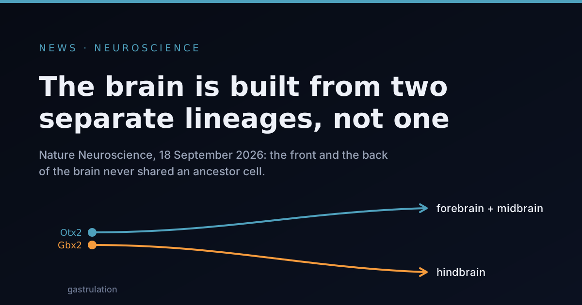
On 18 September, Nature Neuroscience published a paper from a Stanford Medicine group that redraws one of the oldest diagrams in developmental biology. The claim is narrow and checkable: the front of the vertebrate brain and the back of it do not descend from a common ancestor cell. They come from two populations that are already committed to different fates before either one looks like brain tissue, and that never mix.
The diagram being replaced is a tree. In the standard account, the embryo lays down a neural plate, a strip of cells on its back that will roll up into the tube from which the entire nervous system grows. Regional identity arrives afterwards, imposed on an initially uniform sheet by chemical gradients: signals diffusing from organising centres tell one end to become forebrain and the other to become spinal cord. One origin, then specialisation. That framework has organised a century of experiments, and it is the reason a laboratory recipe for brain tissue normally starts by making a generic neural precursor and then pushing it in a direction.
What the Stanford group reports, with Kyle Loh as senior author and the graduate students Carolyn Dundes and Rayyan Jokhai as co-first authors, is two tracks running in parallel rather than one trunk with branches. Working in mouse embryos during gastrulation, the few hours in which a ball of cells sorts itself into the layers that will become every organ, they found two distinct progenitor populations. A progenitor is a cell that still divides and has not finished choosing what it will become. One population expresses Otx2, a gene coding for a transcription factor, a protein whose job is to switch other genes on and off; those cells go on to build the forebrain and midbrain. The other expresses Gbx2, and builds the hindbrain, the structure at the base of the brain that runs breathing, swallowing and facial movement. The two are mutually exclusive from the moment they can be told apart. Neither is the parent of the other.
The supporting evidence sits in the chromatin, the packaging that winds DNA around proteins and decides which stretches are physically available to be read. The anterior and posterior populations, the group reports, carry fundamentally different chromatin configurations. That is a stronger statement than a difference in which genes happen to be active at a given hour. It says each population has already closed off the other’s programme at the level of what the cell can read at all, which is what commitment is supposed to mean, and what a tree model says should not have happened yet.
A cell type nobody could make
The part of the paper likely to matter soonest is not the diagram. It is a cell type.
Human pluripotent stem cells, cells capable of becoming any tissue in the body, have been coaxed into cortical neurons and spinal motor neurons for years. Hindbrain motor neurons, the ones driving the muscles of the face, throat and tongue, have stubbornly resisted. The group’s argument is that everyone was starting in the wrong place: a protocol built on the tree model makes an anterior-type precursor and then tries to push it backwards, which, if the two lineages never connect, is a route that does not exist. Starting instead from a Gbx2 committed posterior state, they report human neurons that fire action potentials, the electrical spikes neurons use to signal, and that carry the protein markers identifying specific hindbrain segments.
That matters because brainstem tissue cannot be taken from living people. Two diseases attack these neurons early: spinal muscular atrophy, and the bulbar onset form of amyotrophic lateral sclerosis, in which speech and swallowing fail before the limbs do. The cells that die first in those patients have never been available in a dish. If the protocol holds up in other hands, that changes.
What the data do not show
The wider claim is the one drawing headlines, and it is the softest part of the story. The two lineage pattern, according to the Stanford announcement, turns up in chickens, zebrafish and acorn worms, marine animals whose lineage separated from ours more than 550 million years ago, as well as in primates. Jellyfish, which diverged earlier still, carry two separate nerve nets at opposite ends of the body. Loh’s framing is that evolution took two pre-existing neural systems and pushed them together in space, so that what looks like a single organ is really a merger.
Read that carefully, because the data underwrite the first half of it and not the second. A pattern conserved across 550 million years of divergence establishes that the arrangement is ancient. It does not establish that two independent nervous systems fused, which is a historical narrative with no fossil, no intermediate form and no experiment behind it. The observation is consistent with the merger story. It is equally consistent with a single ancient nervous system that split its developmental control early and then kept the split.
The rest of the caution is ordinary but worth stating. The lineage work is chiefly mouse embryology; the human contribution is stem cells in culture, not human embryos, so the claim about the human brain is an inference from a conserved mechanism rather than a direct observation. The chromatin result is a correlation between packaging state and eventual fate, not a demonstration that the packaging enforces the fate. And the explanation offered for why earlier hindbrain protocols failed, that they were fighting a lineage barrier, is a hypothesis that fits the outcome, not a controlled comparison. Neurons with the right markers and the right electrical behaviour are the starting point for a disease model, not a disease model.
The two organs phrasing deserves a flag of its own: it is interpretation, not measurement. The adult brain remains one continuous, densely interconnected structure, and nobody thought otherwise on the strength of shared cell ancestry. What actually changes is narrower, and more useful. If regional identity in the nervous system is assigned earlier than assumed, and by lineage rather than by gradients acting on a blank sheet, then the standard recipe for making a given neuron, build something generic and then steer it, is the wrong shape for at least part of the brain. Some cell types will be reachable only by choosing the right track at the start, before there is anything recognisable to steer.
That is a hard constraint on the project of building nervous tissue to order, and it arrived attached to a working example.
Further reading
- Two parallel neural ectoderm progenitors contribute to the developing brain, the paper itself, in Nature Neuroscience, 18 September 2026.
- Human brain is two separate organs, Stanford Medicine-led research finds, the institutional announcement, with the authors’ own framing.
- Two-organ view of the human brain emerges, coverage with more detail on the differentiation protocol.
- The brain is two separate organs joined by evolution, a summary of the cross-species comparisons.